

Case Study Sharing
Case sharing

Staying committed to innovation-driven development, Jiutouniao Medical has been awarded the Second Prize of the Hubei Province Science and Technology Progress Award.
Release date:
2023-04-12 15:44
Source:
On the morning of March 16, the Hubei Provincial Science and Technology Innovation Conference was held at Hongshan Auditorium in Wuhan. During the event, the "Decision of the People's Government of Hubei Province on the 2022 Hubei Provincial Science and Technology Awards" was announced. Among the winners, several groundbreaking achievements from Jiutouniao Medical were honored with the Hubei Provincial Science and Technology Progress Award. Today’s moment of glory belongs to the dedicated science and technology professionals who have tirelessly worked on Hubei’s vibrant land, collectively illuminating the region with the radiant glow of scientific innovation.
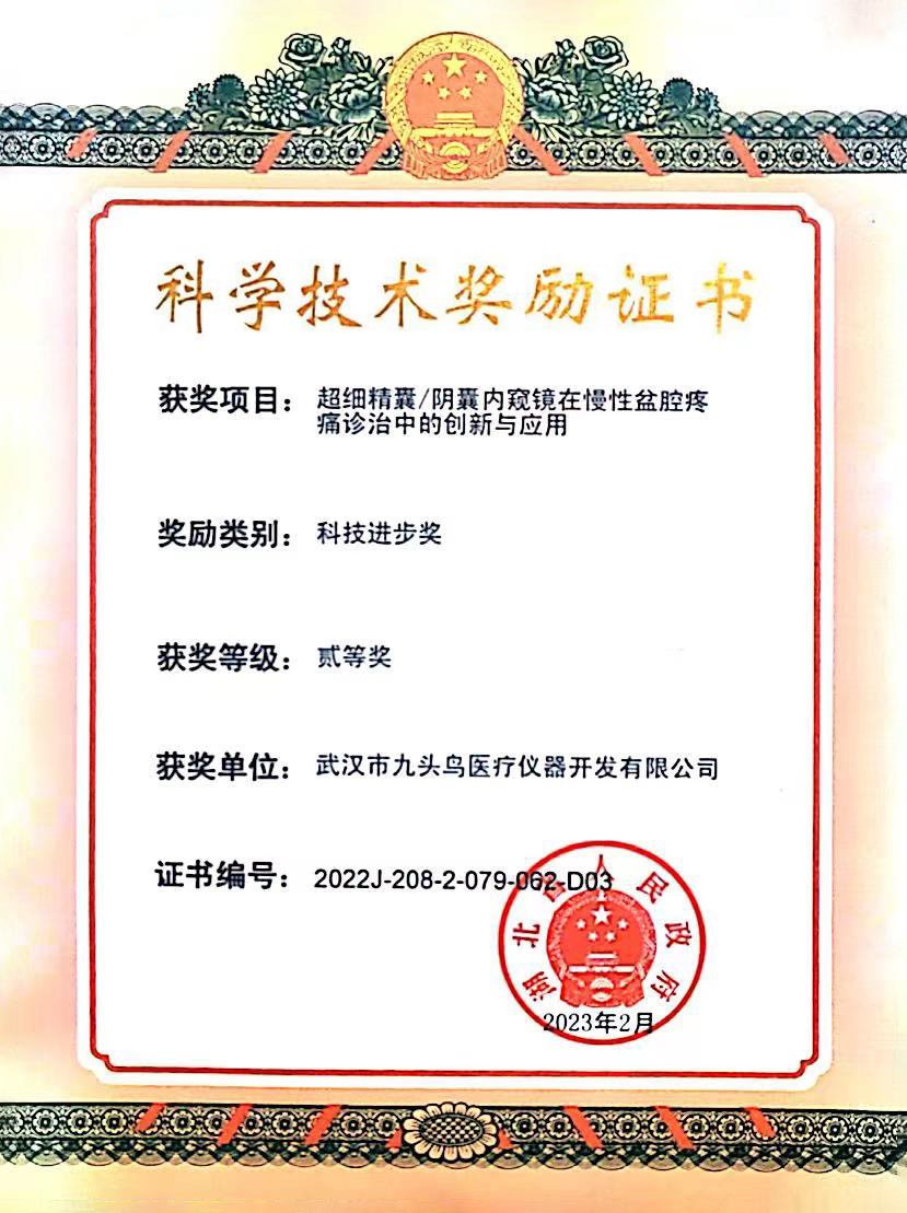
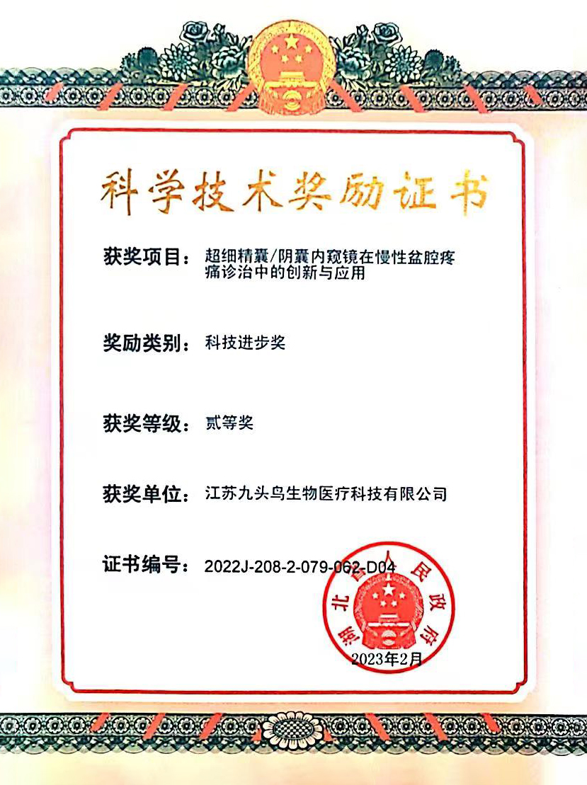
Participated in by Wuhan Jiutouniao Medical Instrument Development Co., Ltd., Wuhan University Zhongnan Hospital Su Xinjun Professor To take the lead of the Ultra-fine seminal vesicle /Innovative Use of Scrotal Endoscopy in the Diagnosis and Treatment of Chronic Pelvic Pain "Applied to Applications" Wins Hubei Province Award Technological Advancement II First prize , this is Among the past award results, Nine-Headed Bird Doctor Achieved the best-ever result in history; at the same time, it is also Nine-headed bird First-time recipient of Hubei Province Science and Technology Progress Award , marking another milestone in the award categories!

Traditionally, diagnosing and treating seminal vesiculitis often requires borrowing instruments like ureteroscopes due to the lack of specialized surgical tools. However, these bulky instruments can only pass naturally through the urethral opening—not through the ejaculatory duct into the seminal vesicles. As a result, clinicians typically resort to methods that disrupt the tissue structure of the ejaculatory duct wall. Alternatively, they may forcefully create an opening to access the seminal vesicles—though this approach is generally less preferred. "The surgical technique of inserting the scope through the prostatic utricle and then entering via a distal incision of the ejaculatory duct disrupts the duct's natural anatomical structure as well as its anti-reflux mechanism at the duct's terminal end, significantly increasing the risk of contamination." The fluid flows back into the vas deferens, Risk of surgical complications such as secondary seminal vesiculitis and testicular epididymitis , but instead will even more likely provoke CPPS Our newly developed ultra-fine seminal vesiculoscope features a thinner diameter compared to similar products, while offering a larger internal working space. Additionally, its precision in visualization is unmatched. 0 °, no refractive angle; the other alternative mirrors used are concave-convex lenses, all of which still have 5-10 Due to the visual difference of °, our developed product causes less damage to the ejaculatory duct during treatment, more effectively preserving its physiological structure. It achieves integrated functions such as real-time display, imaging, and operation, while also enabling more precise, clear, and convenient diagnosis and treatment of seminal vesicle lesions.
At the same time, we are involved. Research and Development & Construction Φ 5mm The ultra-thin scrotal endoscope sheath is inserted into the tunica vaginalis cavity, enabling the ultra-thin scrotal endoscope to perform diagnostic and therapeutic procedures. By establishing a stable operative channel for the scrotal endoscope that remains superficial to the parietal layer of the tunica vaginalis, all other layers of scrotal tissue are carefully shielded from direct communication with the cavity. This ensures that any irrigation fluid used during surgery—when the cavity is filled—cannot inadvertently leak into the scrotal wall. As a result, this innovative approach resolves critical clinical challenges, such as severe postoperative scrotal wall edema and reduced physiological space within the tunica vaginalis cavity, which previously made minimally invasive treatment impossible. Ultimately, this breakthrough overcomes the longstanding industry barrier that has prevented effective, minimally invasive management of scrotal diseases. 。
"With winds rising and waves surging, there will surely be a time to break through—set sail high on cloud-like sails to cross the vast ocean. JiuTouNiao Medical Group will remain steadfast in its commitment to R&D innovation, delivering outstanding clinical equipment to empower healthcare professionals."
Contact us
400-8817-099
-
Address: 9F, Building A1, National Geospatial Information Industrialization Base, 6 Beidou Road, East Lake New Technology Development Zone, Wuhan City
Quick Navigation
Official Official Account
Official Official Account
Internet Drug Information Service License Number: (E)-Non-operational-2020-0044 Wuhan Jiutouniao Medical Instrument Development Co., Ltd. All rights reserved.